
The author (back) and Carly Ebben prepare to collect aerosols on microscopy substrates
Many of us can remember looking through a microscope at a young age and being amazed. All sorts of objects and biological “critters” jumped to life when tiny images that were blown up that we had no clue existed when viewing with the naked eye. This feeling is no doubt similar to what physicist Robert Hooke felt in 1665 when he first identified cells with a microscope. At the University of Iowa we use state-of-the-art microscopes and spectroscopic measurements to analyze aerosols at magnifications far beyond what Robert Hooke could have imagined, and we feel a similar sense of awe as we observe the structure and properties of aerosols with these extremely powerful and sensitive techniques.

A scanning electron microscope (SEM) image of a salt particle generated in our ocean-atmosphere chamber this summer.
The microscopy and spectroscopy measurements that we specialize in in the laboratory of Prof. Vicki Grassian at the University of Iowa are providing a unique view of the aerosols being generated during the intensive measurements at CAICE. Techniques such as transmission electron microscopy (TEM), scanning electron microscopy (SEM), energy dispersive x-ray spectrometry (EDX), Raman microscopy, atomic force microscopy, and x-ray photoelectron microscopy, are allowing use to probe the chemical and physical properties of these marine aerosols in extraordinary detail. For example, our TEM measurements can observe details on the scale of less than 1 nm (for reference that is 40,000 times smaller the diameter of a human hair). The black and what image of a particle from preliminary measurements this summer shows the fine detail that can be obtained for our marine particles (in this case a sea spray particle).
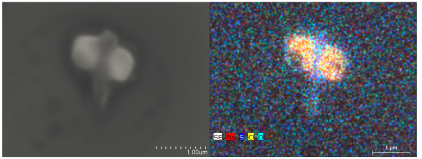
In addition to information on the structure of sea spray particles, we can also learn about the chemical composition from EDX combined with both SEM and TEM. The image on the left shows an image of a particle from SEM and on the right we have mapped out the chemical composition of different features with EDX. From this we can see that a sea salt particle isn’t just NaCl, but that the rods and other features contain mixed cation sulfates. This structure of primary NaCl cube and rods of sulfate species has been shown previously by Wise et al. 2007 for samples of resuspended sea water and field collected aerosols. The similarity of our wave flume generated sea spray during background measurements with previous field measurements shows that we are recreating the ocean well in the wave flume. With the background looking good we are excited to see what the biological species we are adding do to the chemical composition of the aerosols we are seeing!

At left, a traditional image of a salt particle as observed using electron microscopy. At right, the same image, with an overlay of chemical composition obtained using an x-ray spectroscopy technique.
Beyond electron microscopy, we are also excited to be working with ambient characterization techniques that don’t rely on introducing the aerosols to vacuum. Confocal Raman microscopy is one such tool we are utilizing to study aerosol properties as they exist in the atmosphere. The Raman spectra will give us information on organic carbon, sulfate, and other species with characteristic vibrations. Taken together the detailed morphology and chemistry of single particles from microscopy, Raman, and other will provide a great complement to many of the online measurements, in particular single particle size and chemistry from the aerosol time-of-flight mass spectrometer (ATOFMS).
Of course, this is just a sneak peek of the analysis we will be doing on aerosols from the wave flume. Expect more exciting results as soon as we get the samples back to Iowa!
Andrew Ault, Postdoctoral Scholar, Department of Chemistry, University of Iowa